Imaging Gases in Rainbow Colors
—Development of a Pressure-Sensitive Device Capable of Characterizing Gases Using Structural Colors—
2022.11.28
National Institute for Materials Science (NIMS)
NIMS, Harvard University and the University of Connecticut have designed and fabricated a simple device capable of imaging a gas injected into it in multiple colors in accordance with its gaseous properties, enabling chromatic discrimination of different gases.
Abstract
- NIMS, Harvard University and the University of Connecticut have designed and fabricated a simple device capable of imaging a gas injected into it in multiple colors in accordance with its gaseous properties, enabling chromatic discrimination of different gases. This user-friendly device converts the pressure generated by an injected gas into structural colors, thereby imaging it. This technology may potentially have a wide range of applications, such as environmental monitoring, safety assurance and healthcare.
- Imaging of gases is important in many gas-related basic and applied research projects as almost all ambient gases are colorless and invisible. Only a few methods for imaging ambient gas flow have been developed (e.g., the use of infrared cameras capable of detecting temperature changes and airflow measurements by means of releasing tracer particles into the air). These methods require elaborate equipment and are unsuitable for imaging different types of gases in a consistent manner. In addition, the images they produce are unfit for the analysis of gaseous characteristics. A simple method capable of imaging and analyzing all types of gases may have a wide variety of applications, such as image-based measurements.
- This research team recently fabricated a device capable of imaging and differentiating various gases using a wide range of colors (i.e., structural colors) through a simple procedure: polydimethylsiloxane (PDMS)—a soft material—was first shaped into a slab. Part of the PDMS surface was then treated with argon plasma. The plasma-treated PDMS slab was placed on the surface of a glass substrate with its plasma-treated surface down, and they came into complete contact. The plasma-treated PDMS surface forms a periodic ripple-like micropattern when compressed by an injected gas passing through the tight boundary between the PDMS and glass layers. This compression and resultant micropattern formation lead to the production of structural colors. This mechanism is applicable to the imaging and differentiation of any type of gas. When the incoming gas flow is discontinued, structural colors disappear completely. The degree of PDMS deformation depends on the flow rates, viscosities and densities of injected gases. As all gases have unique viscosities and densities, this device can be used to differentiate and analyze gas samples based on these properties under a constant flow rate.
- In future research, the team will work to optimize the device by improving its sensitivity with the goal of making it compatible with various applications (e.g., identification of ambient gases and biological samples). The team will also consider developing a new gas identification technique by combining it with image recognition and machine learning techniques and fabricating a small, CCD (charge coupled device)-integrated device with a simple structure.
- This project was carried out by a research team consisting of Kota Shiba (Senior Researcher, Olfactory Sensors Group, Research Center for Functional Materials, NIMS), David A. Weitz (Professor, Harvard University) and Luyi Sun (Professor, University of Connecticut).
- This research was published in Advanced Science, an open-access journal, on November 17, 2022, local time.
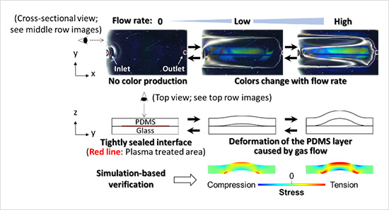
Figure.Mechanism behind the multi-color imaging of a gas injected into the device. Top (top row) and cross-sectional (middle and bottom rows) views of the device and a gas flowing through it.
Related files
- Kota Shiba | Experimental Soft Condensed Matter Group,Harvard University (Open in a new window)
- Research Center for Functional Materials
Contact information
(Regarding this research)
-
Kota Shiba
Senior Researcher
Olfactory Sensors Group
Research Center for Functional Materials
National Institute for Materials Science
Tel: +81-29-860-4603
E-Mail: SHIBA.Kota=nims.go.jp
(Please change "=" to "@")
(General information)
-
Public Relations Office
National Institute for Materials Science
Tel: +81-29-859-2026
Fax: +81-29-859-2017
E-Mail: pressrelease=ml.nims.go.jp
(Please change "=" to "@")
Same Keywords
-
Development of a Novel Mass Analysis Technique That Can Be Performed Even with an Ordinary Business Card
(sensor,device)
2016.08.31
-
Low-Resistance Buffer Layer That Paves the Way for Vertical GaN Devices on Silicon
(device)
2026.06.15
-
Development of a Simple, Revolutionary Printing Technique for Periodic Nano/Microstructures
(polydimethylsiloxane)
2024.09.05