Development of Electron Microscopy Capable of Visualizing Magnetic Structures at the Sub-Nanoscale
—New Technique May Facilitate Research and Development of Next-Generation Spintronic Devices—
2017.10.12
National Institute for Materials Sciences (NIMS)
A NIMS research team developed the world’s first high-resolution Lorentz microscopy capable of observing magnetic structures in materials at sub-nanometer resolutions. The technique enabled the team to visualize magnetic solitons and nanoscale magnetic phase separation in dysprosium, a rare-earth metal.
Abstract
- A NIMS research team developed the world’s first high-resolution Lorentz microscopy capable of observing magnetic structures in materials at sub-nanometer resolutions. The technique enabled the team to visualize magnetic solitons and nanoscale magnetic phase separation in the rare-earth metal dysprosium. The high-resolution magnetic imaging technique developed in this study may accelerate R&D on next-generation magnetic materials.
- Rapid progress has been made in recent years on research and development related to next-generation magnetic materials—particularly spintronic materials. Technologies capable of visualizing nanoscale changes in magnetic fields are vital in these R&D efforts. Lorentz transmission electron microscopy is a promising magnetic imaging technique which would enable direct observation of such phenomena. However, large spherical and chromatic aberrations of unique electron lenses (i.e., Lorentz lenses) used in this microscopy had limited resolution to approximately 2 to 10 nm. Spatial resolution down to 1 nm or less is necessary to accurately observe nanoscale magnetic structures.
- A NIMS research team succeeded in developing the world’s first Lorentz microscopy with sub-nanometer spatial resolution (0.6 nm or higher). This was achieved by simultaneously using a spherical aberration corrector and an electron beam monochromator, which greatly reduced the high-order aberrations (e.g., spherical aberration) and chromatic aberration of the Lorentz lens. It had been suggested that multiple magnetic phases exist in dysprosium, and that these magnetic phases are potentially applicable to spintronics. The team observed these phases using the new microscopic technique and found that magnetic solitons exist in dysprosium in the absence of an external magnetic field. In addition, the team succeeded in visualizing nanoscale magnetic phase separation induced by external magnetic fields. We expect these observations to serve as vital fundamental knowledge for spintronics research.
- Active efforts have been made in next-generation spintronic device R&D to produce novel functional properties by controlling the nanoscale magnetic structures formed in magnetic bodies. We expect that Lorentz microscopy capable of visualizing magnetic structures at the sub-nanoscale will expedite R&D efforts.
- This research was carried out by a NIMS research team led by Senior Engineer Takuro Nagai (of both the Electron Microscopy Group and the Transmission Electron Microscopy Station of the Research Network and Facility Services Division), Koji Kimoto (Deputy Director-General of the Research Center for Advanced Measurement and Characterization) and Masaki Takeguchi (Director of the Transmission Electron Microscopy Station). Part of this study was conducted jointly with Koji Inoke (Senior Research Specialist, FEI Company Japan Ltd. (currently affiliated with Nippon Roper K.K.)) and funded by a JSPS Grant-in-Aid for Scientific Research (Project Number: JP17K04984) and the MEXT Nanotechnology Platform Japan program.
- This study was published in the online version of Physical Review B (in its Rapid Communications section), a journal of the American Physical Society, as an Editors’ Suggestion article on September 19, 2017, local time.
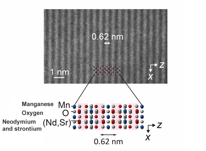
Figure : An electron microscope image of a layered manganese oxide, Nd0.2Sr1.8MnO4, observed using the newly developed technique. The 0.62 nm lattice period can be recognized in this lattice image.
Related files
- Research Center for Advanced Measurement and Characterization
- Transmission Electron Microscopy Station
Contacts
(Regarding this research)
-
Takuro Nagai,
Senior Engineer,
Transmission Electron Microscopy Station, High Resolution Group,
Research Network and Facility Services Division,
National Institute for Materials Science (NIMS)
Tel: +81-29-859-2132
E-Mail: NAGAI.Takuro=nims.go.jp
(Please change "=" to "@")
(For general inquiries)
-
Public Relations Office, Planning Division
National Institute for Materials Science
1-2-1 Sengen, Tsukuba, Ibaraki, 305-0047, Japan
Tel: +81-29-859-2026
Fax: +81-29-859-2017
E-Mail: pressrelease=ml.nims.go.jp
(Please change "=" to "@")
Recent Press Release
-
No Need for Rare Earths or Liquid Helium! Cryogenic Cooling Material Composed Solely of Abundant Elements
2025.12.24
-
Novel Materials Design Approach Achieves a Giant Cooling Effect and Excellent Durability in Magnetic Refrigeration Materials
2025.12.19
-
Multiple Autonomous AI Systems Spontaneously Collaborate to Advance Materials Research
2025.12.10