Cutting-edge Nanostructure Analysis using Atom Probe Tomography (APT)
APT
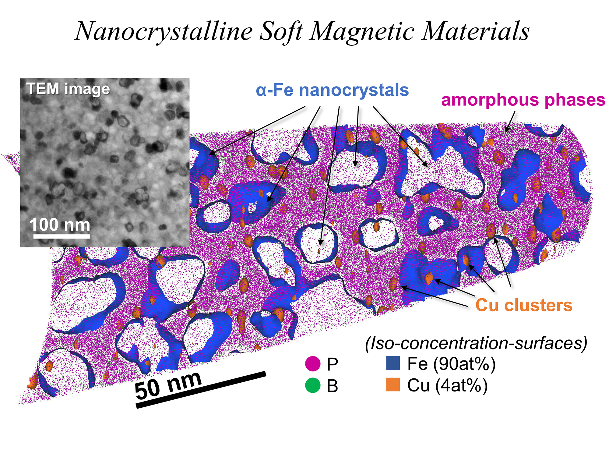
Atom probe tomography (APT, or 3DAP; three-dimentional atom probe) is the only method to visualize the distribution of atoms as 3D map at a magnification ratio of over 1 million by simultaneously measuring the mass and position of atoms ionized with laser pulses (or voltage pulses) from the needle-shaped specimens with the radius of curvature ~ 50 nm. For example, this method can precisely analyze the 3D distribution of elements within nanoscale devices or the uneven elemental distributions within materials. The development of laser-assisted atom probe tomography (APT) in the early 2000s has enabled the three-dimensional elucidation of the distribution of trace elements and dopants at the atomic level, not only in metallic materials but also in semiconductors and insulators.
APT
×TEM
Our unit focuses on the APT method, performing complementary analysis combined with transmission electron microscopy (TEM) while collaborating with Electron Microscopy Unit since the crystal structure or types of defects must be determined by using TEM technique. This allows us to provide sophisticated and advanced nanostructure analysis. We also provide comprehensive technical support from scanning electron microscopy (SEM) observation to APT specimens preparation by using focused ion beam (FIB).
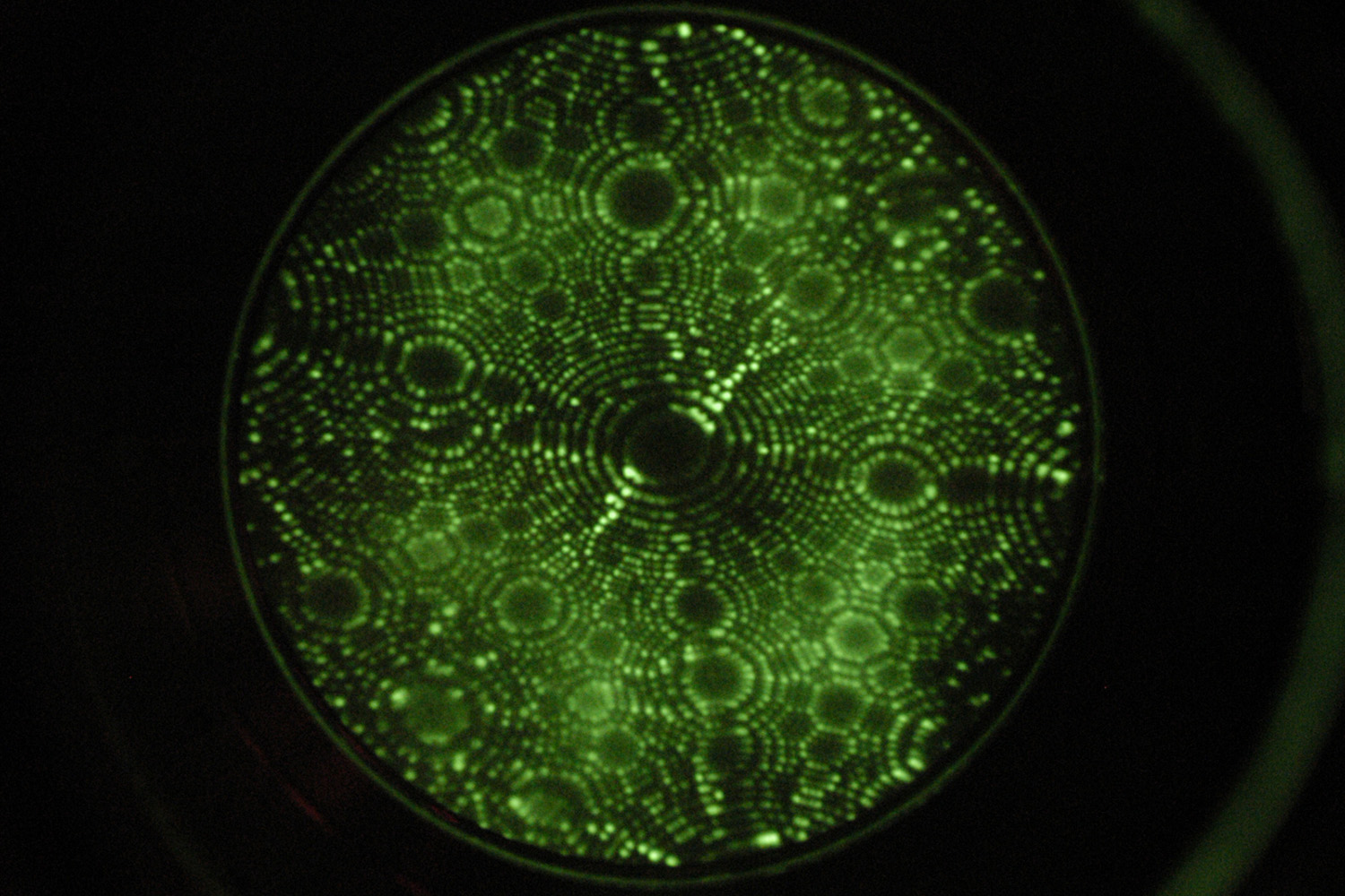
FIM
APT is based on the "Field Ion Microscopy (FIM)" technique, which, in 1951, enabled humanity to directly observe atoms for the first time. In this technique, a needle-shaped specimen is cooled to several tens of kelvin in a vacuum chamber and a positive voltage is applied. When an imaging gas such as He or Ne is introduced into the chamber, the gas atoms are attracted to the surface of the needle-shaped specimen, which is under a high electric field. The imaging gas atoms (ions), which have lost their thermal (kinetic) energy due to cooling, are then accelerated by the electric field between the specimen surface and a fluorescent screen, ultimately striking the fluorescent screen and producing observable bright spots. Furthermore, as the voltage applied to the needle-shaped specimen is increased, "Field Evaporation" occurs, whereby the atoms on the surface of the needle-shaped specimen themselves are ionized.
Skill

Jun Uzuhashi (Unit Leader)
E-mail: UZUHASHI.Jun[at]nims.go.jp
View Jun Uzuhashi's Profile (SAMURAI)
Kyoko Suzuki (Project Engineer)
E-mail: SUZUKI.Kyoko[at]nims.go.jp
supported by 3 Office Staffs