Techniques for Elemental Analysis and Imaging Using X-Ray Measurements Obtained from an Ordinary Digital Camera
—The Simple Method Promotes X-Ray Analysis Anywhere and Anytime—
National Institute for Materials Science (NIMS)
A NIMS research group succeeded in developing new techniques to perform analysis and imaging of chemical elements by taking images of a target material using an ordinary, visible-light digital camera with a slight modification, and obtaining X-ray spectra from processed images.
(“Seeing elements by visible-light digital camera”, Wenyang Zhao &
Kenji Sakurai, Scientific Reports 7, Article number: 45472 (2017), doi:
10.1038/srep45472)
Abstract
- Kenji Sakurai and Wenyang Zhao, managing researcher and junior researcher, respectively, X-Ray Physics Group, RCAMC, NIMS, succeeded in developing new techniques to perform analysis and imaging of chemical elements by taking images of a target material using an ordinary, visible-light digital camera with a slight modification, and obtaining X-ray spectra from processed images. The technique is expected to allow simpler and convenient X-ray analysis applicable to a wider range of fields under diverse conditions.
- Materials consist of various chemical elements, and their physical/chemical properties are greatly influenced by their compositions. Therefore, it is important to perform qualitative and quantitative analysis of chemical elements composing a material for deeper understanding of the material and the development of new materials. It is known that the types and amounts of chemical elements present in a material can be determined by irradiating the material with X-rays and measuring the energy levels and intensities of the fluorescent X-rays emitted from the material in response to the irradiation. Currently, the analysis of fluorescent X-rays requires the use of an X-ray fluorescence spectrometer or X-ray detector. In addition, to investigate the distribution of different elements across a sample, it is necessary to use a more expensive detector.
- The research team recently discovered methods to conduct analysis and imaging of chemical elements using fluorescent X-ray data obtained from a digital camera equipped with a visible-light CMOS (complementary-metal-oxide-semiconductor) device, which is often incorporated in optical microscopes. The camera was also equipped with an opaque and thin window, which only allows X-rays to penetrate, between the lens and sensor. When fluorescent X-rays emitted from a sample pass through the window and enter the CMOS device, electric charges are generated. The energy of incident X-rays can be quantified by instantaneously measuring the amount of charges generated. However, charges generated are recorded across multiple pixels, and pixel data is frequently lost. To address this issue, the team developed a method to retrieve the lost data by examining the dispersion state of charges across pixels and by processing images for both the amount and position of charges initially recorded. Using this method, the team was able to obtain reliable X-ray spectra in a stable manner. The team analyzed fluorescent X-rays emitted from a ceramic plate shown in the figure below using the developed method, and detected cobalt only in the upper plate surface which was painted blue, but not in the bottom surface.
- The research team also succeeded in creating images showing the distribution of different elements across a sample, using the pinhole camera principle. In future studies, the team hopes to contribute to materials development by applying the techniques to visualize the movement of chemical elements in video images and trace chemical reaction processes.
- This study (doi:10.1038/srep45472) was published in Springer Nature's online journal, Scientific Reports, on March 31, 2017, local time.
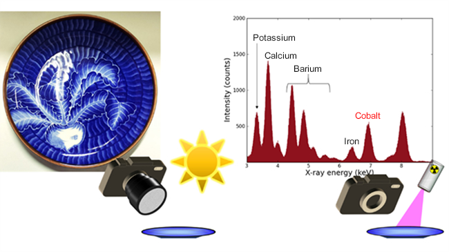
Figure. (Left) A ceramic plate used as a measurement sample. Use of a visible light camera allows one to record target objects’ shapes and colors as images (The camera used in this study takes black and white images). (Right) X-ray spectra obtained using the same camera. Chemical elements can be analyzed based on differences in energy levels, which represent different X-ray colors, distributed across the X-ray wavelength region.