近年、集束イオンビーム(FIB)-走査型電子顕微鏡(SEM)複合装置による微細加工技術が発展したことにより、結晶粒界、異相界面、多層膜、デバイスの任意領域、ナノワイヤ等から3DAP測定用試料を作製して、3DAP分析することが可能となってきています。
このページでは、NIMSでこれまでに実施してきた3DAPによる分析例の一部を紹介しています。ページ下部には
論文リストの一部も掲載しています。
3次元アトムプローブ(3DAP)を用いた分析例
ネオジム磁石の結晶粒界
ネオジム磁石(Nd-Fe-B)の結晶粒界の3次元原子マップです。3DAP分析により、微量に添加したGaやCuが結晶粒界に偏析している様子が確認できます。
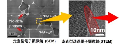
ネオジム磁石断面のSEM像(左)
結晶粒界の高倍率STEM像(右)
(● Nd ● Ga ● Cu)
3DAPによる結晶粒界の3次元原子マップ~35×35×110 nm3
(参考:
Broadening the applications of the atom probe technique by ultraviolet femtosecond laser, K. Hono et al.,
Ultramicroscopy 111, 576 (2011).)
マグネシウム合金の転移線への合金元素の偏析
マグネシウム合金において合金元素(Ca, Zn)が転位線へ偏析している様子をTEMと3DAPの両方を用いて解析した結果です。
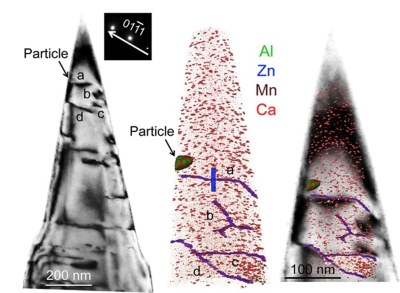
3DAP分析前に3DAP測定用試料そのものをTEM観察し転移線の位置を確認し(左)、3DAP測定により得られた3次元原子マップ(中)をTEM像と重ね合わせることにより(右)、転移線に沿って合金元素が偏析している様子が確認できます。
(参考:
Bake-hardenable Mg-Al-Zn-Mn-Ca sheet alloy processed by twin-roll casting, M.Z. Bian et al.,
Acta Mater. 158, 278-288 (2018).)
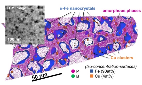
ナノ結晶軟磁性材料の3次元原子マップ(スライス)
3次元アトムプローブではTEM試料の膜厚よりも小さな構造物(数nm~数十nm)の元素分布を精緻に解析することが可能です。その一例としてナノ結晶軟磁性材料が挙げられます。
(参考:
Heating rate dependence of coercivity and microstructure of Fe-B-P-Cu nanocrystalline soft magnetic materials, Y. Nomura et al.,
J. Alloys Compd. 859, 157832 (2021).)
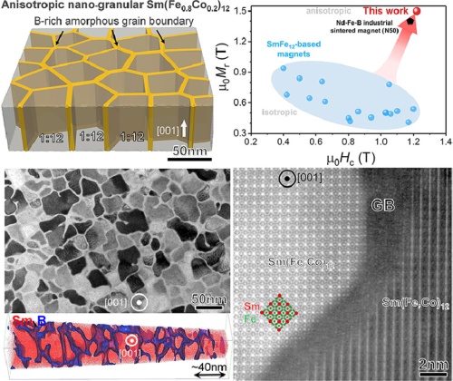
ホウ素添加サマリウム鉄コバルト化合物薄膜のナノ組織解析
厚さ約3nmのアモルファス相がSm(Fe0.8Co0.2)12粒子を均一に覆うユニークな複相ナノ構造のナノ組織解析例です。薄膜によるモデル実験ですが、サマリウム鉄系磁石がネオジム磁石を超えるポテンシャルを実証しました。
(参考:
Achievement of high coercivity in Sm(Fe0.8Co0.2)12 anisotropic magnetic thin film by boron doping, H. Sepehri-Amin et al.,
Acta Mater. 194, 337-342 (2020).)
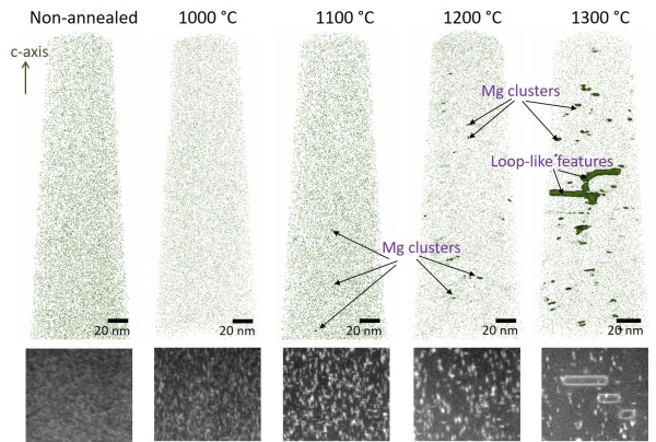
Mgイオン注入GaN(窒化ガリウム)中のMgの3次元原子マップ
p型GaN半導体実現に必要となる微量なMgの分布を3DAPによって可視化した結果です。それぞれのMg原子マップの下部には構造の揺らぎを敏感に検出可能な低角環状暗視野(LAADF)-STEM像を示しています。なおここに示しているGaNにイオン注入されたMg濃度は1×1019cm-3(約0.01at.%)です。
(参考:
Atomic-scale quantitative analysis of implanted Mg in annealed GaN layers on free-standing GaN substrates, A. Kumar et al.,
J. Appl. Phys. 126, 235704 (2019).)
ZrO2-MgAl2O4ナノコンポジットセラミックス(絶縁体バルク材料)
紫外光波長のフェムト秒レーザーを用いたNIMS独自開発3DAPにより、世界で初めて絶縁体バルク材料の解析に成功しました。これは短波長レーザー補助が半導体や絶縁体の解析に有効であることを示す結果で、3DAPの応用範囲拡大に貢献しました。
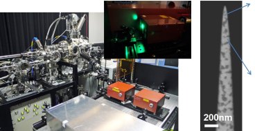
NIMS独自開発レーザー補助3DAP(左)
3DAP測定用試料のSEM像(右)
(● Mg ● Al)
ジルコニア・スピネルナノコンポジットセラミックスの紫外光フェムト秒レーザーを用いた3DAPによる3次元原子マップ ~65×65×270 nm3
(絶縁体バルク材料の世界発の解析例)
(参考:
Laser-assisted atom probe analysis of zirconia/spnel nanocomposite ceramics, Y.M. Chen et al.,
Scr. Mater. 61, 693-696 (2009).)
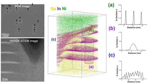
GaN/GaInN:ピット(V型欠陥)直下でのIn分布
STEM観察では原理上、得られる情報は観察試料の膜厚方向に投影したものであるために、膜厚方向に3次元的な組織変化があった場合には、その解析が困難です。そのような場合には3DAP分析を用いることで3次元での詳細な元素分布解析が可能となります。
(参考:
Atomic scale characterization of GaInN/GaN multiple quantum wells in V-shaped pits, S. Tomiya et al.,
Appl. Phys. Lett. 98, 181904 (2011).)
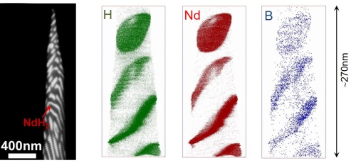
水素化合物の解析例(軽元素の分析も可能)
3DAPは高電圧を印加した試料にレーザーパルス(または電圧パルス)を加えることにより試料表面から原子をイオン化させ、そのイオンの質量を飛行時間により同定する原理(Time-of-Flight Mass Spectrometer)ですので、水素などの軽元素の分析も可能です。
(参考:
Quantitative laser atom probe analyses of hydrogenation-disproportionated Nd-Fe-B powders, H. Sepehri-Amin et al.,
Ultramicroscopy 111, 615 - 618 (2011).)
GaN(窒化ガリウム)ナノワイヤの多層量子井戸構造
FIB-SEM複合装置の発達により数百nm程度の3次元構造物内から任意領域を選んで3DAP分析することも可能です。
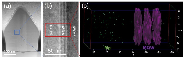
(a) ナノワイヤの形状(HAADF-STEM像)、(b) MQW(多層量子井戸構造)のHAADF-STEM像、(c) 任意領域からの3次元原子マップ
(参考:
Characterizations of GaN nanowires and GaInN/GaN multi-quantum shells grown by MOVPE, N. Goto et al.,
Jpn. J. Appl. Phys. 59, SGGE05 (2020).)
ナノボイドを含むZnO(酸化亜鉛)ナノワイヤ
ナノワイヤのような形状のサンプルであっても工夫次第で3DAP測定用試料の作成が可能です。またこの分析例ではSbドープZnOナノワイヤ中に含まれるナノボイドの可視化にも成功しました。
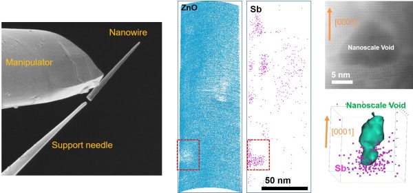
試料の作製方法(左)、スライスした3次元原子マップ(中)、ナノボイドのHAADF-STEM像(右上)
(参考:
Investigation of nanoscale voids in Sb-doped p-type ZnO nanowires, K.C Pradel et al.,
Nanotechnology 29, 335204 (2018).)
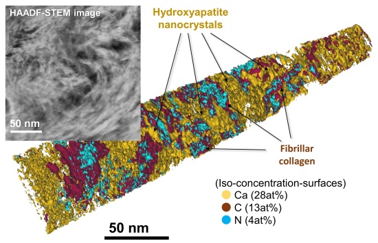
豚の歯(象牙質)
象牙質とは約70%をヒドロキシアパタイトと呼ばれる無機質、約20%を有機物、その他に水分で構成される歯の主体です。有機物を多く含む材料ですが、このような材料の3次元アトムプローブ解析にも成功しています。
(参考:
X-ray diffraction and in situ pressurization of dentine apatite nanocrystals quantifies modulus stiffening upon carbonate removal, J-B. Forien et al.,
Acta Biomater. 120, 91-103 (2021).)
Achievement of high coercivity in Sm(Fe0.8Co0.2)12 anisotropic magnetic thin film by boron doping, H. Sepehri-Amin et al., Acta Mater. 194, 337-342 (2020).
Magnetic anisotropy constants of ThMn12-type Sm(Fe1-xCox)12 compounds and their temperature dependence, D. Ogawa et al., J. Magn. Magn. Mater. 497, 165965 (2020).
Comparison of coercivity and squareness in hot-deformed and sintered magnets produced from a Nd-Fe-B-Cu-Ga alloy, X.D. Xu et al., Scr. Mater. 160, 9-14 (2019).
Role of Ga on the high coercivity of Nd-rich Ga-doped Nd-Fe-B sintered magnet, T.T. Sasaki et al., J. Alloys. Comp. 790, 750-759 (2019).
Microstructure of a Dy-free Nd-Fe-B sintered magnet with 2T coercivity, X.D. Xu et al., Acta Mater. 156, 146-157 (2018).
Suppression of non-oriented grains in Nd-Fe-B hot-deformed magnets by Nb doping, X. Tang et al., Scr. Mater. 147, 108-113 (2018).
Coercivity enhancement of hot-deformed Ce-Fe-B magnets by grain boundary infiltration of Nd-Cu eutectic alloy, X. Tang et al., Acta Mater. 144, 884?895 (2018).
Correlation of microchemistry of cell boundary phase and interface structure to the coercivity of Sm(Co0.784Fe0.100Cu0.088Zr0.028)7.19 sintered magnets, H. Sepehri-Amin et al., Acta Mater. 126, 1-10 (2017).
Effect of carbon on the coercivity and microstructure in fine-grained Nd-Fe-B sintered magnet, T.T. Sasaki et al., Acta Mater. 84, 506-514 (2015).
Grain size dependence of coercivity of hot-deformed Nd-Fe-B anisotropic magnets, J. Liu et al., Acta Mater. 82, 336-343 (2015).
Microstructure evolution of hot-deformed Nd-Fe-B anisotropic magnets, J. Liu et al., J. Appl. Phys. 115, 17A744 (2014).
High-coercivity ultrafine-grained anisotropic Nd-Fe-B magnets processed by hot deformation and the Nd-Cu grain boundary diffusion process, H. Sepehri-Amin et al., Acta Mater. 61, 6622-6634 (2013).
Enhancement of coercivity of hot-deformed Nd-Fe-B anisotropic magnet by low-temperature grain boundary diffusion of Nd60Dy20Cu20 eutectic alloy, H. Sepehri-Amin et al., Scr. Mater. 69, 647-650 (2013).
Effect of Nd content on the microstructure and coercivity of hot-deformed Nd-Fe-B permanent magnets, J. Liu et al., Acta Mater. 61, 5387-5399 (2013).
High coercivity Nd-Fe-B thick films without heavy rare earth additions, N.M. Dempsey et al., Acta Mater. 61, 4920-4927 (2013).
The mechanism of coercivity enhancement by the grain boundary diffusion process of Nd-Fe-B sintered magnets, H. Sepehri-Amin et al., Acta Mater. 61, 1982-1990 (2013).
Correlative multi-scale characterization of a fine grained Nd-Fe-B sintered magnet, T.T. Sasaki et al., Ultramicroscopy 132, 222-226 (2013).
The effect of the thermal decomposition reaction on the mechanical and magnetocaloric properties of La(Fe,Si,Co)13, K. Lowe et al., Acta Mater. 60, 4268-4276 (2012).
Enhanced coercivity of spark plasma sintered Zn-bonded Sm-Fe-N magnets, D. Prabhu et al., Scr. Mater. 67, 153-156 (2012).
Grain boundary and interface chemistry of a Nd-Fe-B based sintered magnet, H. Sepehri-Amin et al., Acta Mater. 60, 819 (2012).
Microstructure of fine grained Nd-Fe-B sintered magnets with high coercivity, H. Sepehri-Amin et al., Scr. Mater. 65, 396-399 (2011).
Distribution of Dy in high-coercivity (Nd,Dy)-Fe-B sintered magnets, W.F. Li et al., Acta Mater. 59, 3061 (2011).
Quantitative laser atom probe analyses of hydrogenation-disproportionated Nd-Fe-B powders, H. Sepehri-Amin et al., Ultramicroscopy 111, 615 - 618 (2011).
「ネオジム磁石の微細構造と保磁力」宝野和博、『ネオジム磁石のすべて』(佐川眞人監修)第7章、アグネ技術センター(2011年)
Coercivity enhancement of hydrogenation-disproportionation-desorption-recombination processed Nd-Fe-B powders by the diffusion of Nd-Cu eutectic alloys, H. Sepehri-Amin et al., Scr. Mater. 63, 1124-1127 (2010).
Grain boundary structure and chemistry of Dy-diffused Nd-Fe-B sintered magnets, H. Sepehri-Amin et al., J. Appl. Phys. 107, 09A745 (2010).
Effect of Ga addition on the microstructure and magnetic properties of hydrogenation-disproportionation-desorption-recombination processed Nd-Fe-B powder, H. Sepehri-Amin et al., Acta Mater. 58, 1309 (2010).
Direct evidence for Cu concentration variation and its correlation to coercivity in Sm(Co0.74Fe0.1Cu0.12Zr0.04)7.4 ribbons, R. Gopalan et al., Scr. Mater. 60, 764-767 (2009).
Effect of post-sinter annealing on the coercivity and microstructure of Nd-Fe-B permanent magnets, W.F. Li et al., Acta Mater. 57, 1337-1346 (2009).
The role of Cu addition in the coercivity enhancement of sintered Nd-Fe-B permanent magnets, W.F. Li et al., J. Mater. Res. 24, 413 (2009).
Heating rate dependence of coercivity and microstructure of Fe-B-P-Cu nanocrystalline soft magnetic materials, Y. Nomura et al., J. Alloys Compd. 859, 157832 (2021).
The effect of Co addition on magnetic and structural properties of nanocrystalline (Fe,Co)-Si-B-P-Cu alloys, M. Kuhnt et al., J. Alloys. Comp. 766, 686-693 (2018).
Three-dimensional atom probe analysis and magnetic properties of Fe85Cu1Si2B8P4 melt spun ribbons, S. Jafari et ai., J. Magn. Magn. Mater. 401, 1123-1129 (2016).
Three-dimensional atom probe study of Fe–B-based nanocrystalline soft magnetic material, Y.M. Chen et al., Acta Mater. 57, 4463 (2009).
Composition and non-equilibrium crystallization in partially devitrified Co-rich soft magnetic nanocomposite alloys, P.R. Ohodnicki et al., Acta Mater. 57, 87-96 (2009).
Simultaneous achievement of high thermal conductivity, high strength and formability in Mg-Zn-Ca-Zr sheet alloy, Z.H. Li et al., Mater. Res. Lett. 8, 335-340 (2020).
Bake-hardenable Mg-Al-Zn-Mn-Ca sheet alloy processed by twin-roll casting, M.Z. Bian et al., Acta Mater. 158, 278-288 (2018).
Precipitation in a Ag-Containing Mg-Y-Zn Alloy, Y.M. Zhu et al., Metall. Mater. Trans. A 47, 927-940 (2016).
Strong and ductile heat-treatable Mg-Sn-Zn-Al wrought alloys, T.T. Sasaki et al., Acta Mater. 99, 176-186 (2015).
The effect of Ag and Ca additions on the age hardening response of Mg-Zn alloys, T. Bhattacharjee et al., Mat. Sci. Eng. A 575, 231 (2013).
Significant enhancement of age hardening response in Mg-10Sn-3Al-1Zn alloy by Na microalloying, F.R. Elsayed et al., Scr. Mater. 68, 797-800 (2013).
Effect of Li additions on the age hardening response and precipitate microstructures of Mg-2.4Zn-0.16Zr based alloys, C.L. Mendis., Mater. Sci. Eng. A 535, 122-128 (2012).
Unexpected influence of Mn addition on the creep property in a cast Mg-2Al-2Ca (mass%) alloy, T. Homma et al., Acta Mater. 59, 7662-7672 (2011).
Quantitative atom probe analyses of magnesium alloys, K. Oh-ishi et al., Ultramicroscopy 111, 715 (2011).
Microstructures and tensile properties of a twin roll cast and heat treated Mg-2.4Zn-0.1Ag-0.1Ca-0.1Zr alloy, C.L. Mendis et al., Scr. Mater. 64, 335-338 (2011).
Enhanced precipitation hardening of Mg-Ca alloy by Al addition, J. Jayaraj, et al., Scr. Mater. 63, 710-715 (2010).
Effect of Al additions on the age hardening response of a Mg-2.4Zn-0.1Ag-0.1Ca (at.%) alloy - TEM and 3DAP study, C.L. Mendis et al., Mat. Sci. Eng. A 527, 973-980 (2010).
Bimodally grained microstructure development during hot extrusion of Mg-2.4Zn-0.1Ag-0.1Ca-0.16Zr (at.%) alloy, K. Oh-ishi et al., Acta Mater. 57, 5593-5604 (2009).
Precipitation-hardenable Mg-2.4Zn-0.1Ag-0.1Ca-0.16Zr (at%) wrought magnesium alloy, C.L. Mendis et al., Acta Mater. 57, 749-760 (2009).
Combined APT and STEM Analyses, A. Kumar and T. Ohkubo, AIP Book, Characterization of Defects and Deep Levels for GaN Power Devices, Chapter 5 (2020).
Mg diffusion and activation along threading dislocations in GaN, W. Yi et al., Appl. Phys. Lett. 116, 242103 (2020).
Atomic-scale quantitative analysis of implanted Mg in annealed GaN layers on free-standing GaN substrates, A. Kumar et al., J. Appl. Phys. 126, 235704 (2019).
Characterizations of GaN nanowires and GaInN/GaN multi-quantum shells grown by MOVPE, N. Goto et al., Jpn. J. Appl. Phys. 59, SGGE05 (2020).
Influence of photoexcited carriers on compositional measurements by APT: AlGaN alloy case study, Y. Kanitani et al., Jpn. J. Appl. Phys. 58, 096505 (2019).
Atomic scale characterization of GaInN/GaN multiple quantum wells in V-shaped pits, S. Tomiya et al., Appl. Phys. Lett. 98, 181904 (2011).
鉄鋼材料
Direct observation of carbon distribution in retained austenite films in low carbon lath martensite steel, S. Morito et al., ISIJ International 51, 1200 (2011).
Bimodally grained high strength Fe fabricated by mechanical alloying and spark plasma sintering, B. Srinivasarao et al., Acta Mater. 57, 3277 (2009).
Complementary use of transmission electron microscopy and atom probe tomography for the investigation of steels nanostructured by severe plastic deformation, X. Sauvage et al., Scr. Mater. 60, 1056-1061 (2009).
Quantitative Analysis of Sulfur Segregation at the Oxide/Substrate Interface in Ni-Base Single Crystal Superalloy, C. Tabata et al., Scr. Mater. 194, 113616 (2021).
Laser-assisted atom probe analysis of zirconia/spnel nanocomposite ceramics, Y.M. Chen et al., Scr. Mater. 61, 693-696 (2009).
X-ray diffraction and in situ pressurization of dentine apatite nanocrystals quantifies modulus stiffening upon carbonate removal, J-B. Forien et al., Acta Biomater. 120, 91-103 (2021).
Face-selective tungstate ions drive zinc oxide nanowire growth direction and dopant incorporation, J. Liu et al., Comms. Mater. 1, 58 (2020).
Quantitative atom probe analyses of rare-eath-doped ceria by femtosecond laser, F. Li et al., Ultramicroscopy 111, 589-594 (2011).
Laser-assisted three dimensional atom probe analysis of dopant distribution in Gd-doped CeO2, F. Li et al., Scr. Mater. 63, 332-335 (2010).
3DAP analysis of (Ga,Mn)As diluted magnetic semiconductor thin film, M. Kodzuka et al., Ultramicroscopy 109, 644-648 (2009).
Investigation of nanoscale voids in Sb-doped p-type ZnO nanowires, K.C Pradel et al., Nanotechnology 29, 335204 (2018).
Laser assisted atom probe analysis of thin films on insulating substrates, M. Kodzuka., Ultramicroscopy 111, 557 (2011).
Evidence for nano-Si clusters in amorphous SiO anode materials for rechargeable Li-ion batteries, H. Sepehri-Amin et al., Scr. Mater. 69, 92 (2013).
Laser assisted field evaporation of oxides in atom probe analysis, Y.M. Chen et al., Ultramicroscopy 111, 562-566 (2011).
「レーザーアトムプローブによるナノ組織解析」大久保忠勝、表面と真空 61(特集「ナノスケール3次元分析の最前線」)pp 778-783 (2018年).
Atom probe tomography of metallic nanostructures, K. Hono et al., MRS Bulletin 41, Atom Probe Tomography, 23-29 (2016).
Broadening the applications of the atom probe technique by ultraviolet femtosecond laser, K. Hono et al., Ultramicroscopy 111, 576 (2011).
Mechanism of laser assisted field evaporation from insulating oxides, M. Tsukada et al., Ultramicroscopy 111, 567-570 (2011).
Influence of the wavelength on the spatial resolution of pulsed-laser atom probe, B. Gault et al., J. Appl. Phys. 110, 094901 (2011).
Influence of laser irradiation condition on a femtosecond laser assisted tomographic atom probe, A. Nishimura et al., Ultramicroscopy 109, 467-471 (2009).