Functionalization of Sea Urchin Skeletons
Currently, sea urchins are highly valued as a food ingredient both in Japan and overseas, and are caught not only in Japan but also in China, Chile, Russia, the United States, Canada, and other countries. However, the edible parts, the gonads, only account for around 20% by mass, and the rest are discarded, resulting in disposal costs.
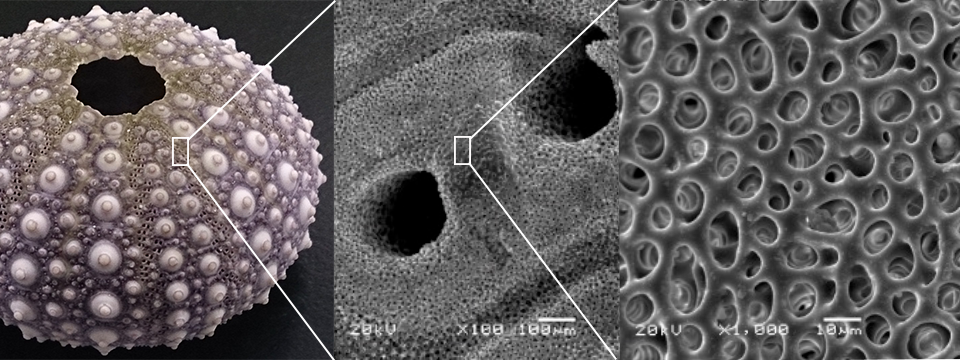
Scanning electron microscopic observation of sea urchin skeltons illustrates pores of several hundred µm and pores of a dozen µm, as shown in the photo above. Such porous structure is difficult to create artificially. So, development of functional materials by utilizing this pore structure created by nature leads to the reuse of waste materials. This make possible to be sold (or at least to be free salvaged) as valuable resources, instead of paying disposal fees, and reduce the economic burden of sea urchin fishing.
Further, sea urchins eat a lot of seaweed and be said to cause coastal denudation; thus, Inedible sea urchins are sometimes exterminated. Even though, their waste can also be used in the same way.
In the past, some people try to use extracted sea urchin skeletons as filters for aquariums. Currently, we have demonstrated that simple treatment of sea urchin skeletons can lead to a variety of uses.
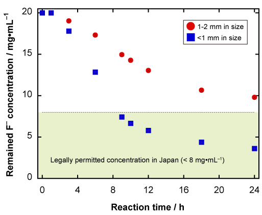
The figure above shows fluoride ion removal amounts as a fucntion of time by phosphatized sea urchin skeleton granules. Granules smaller than 1 mm removed fluoride ions to become a value less than discharging regulation in 9 hours. Furthermore, with this method, the relatively small apatite granules containing fluoride ions are automatically released from the original relatively large granules and can be easily separated from each other. Therefore, allowing fluoride ions and phosphate ions to be recovered simultaneously as apatite granules.
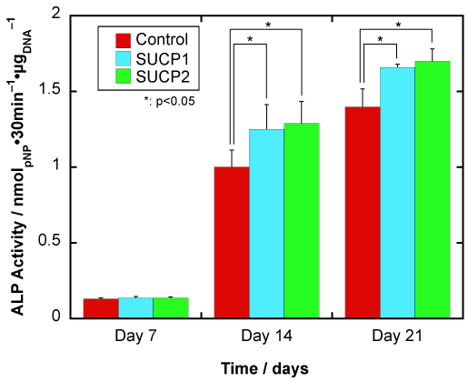
On the other hand, the left graph shows the results of a cell culture test evaluating bone formation of a bone regeneration scaffold, which can also be used as a bone void fillers, composed of phosphatized sea urchin skeleton granules and gelatin. The bone regeneration function was evaluated by production amounts of alkaline phosphatase (ALP), one of the proteins that indicates the bone formation function of osteoblasts.
The scaffold materials using sea urchin skeletal granules (SUCP1, SUCP2) produced significantly more ALP on days 14 and 21 of culture compared to the control gelatin sponge group, indicating improved bone formation function of the cells.
Gene expression measurements for the ALP and osteocalcin (another protein that can estimate bone formation function) also indicated higher on the 14th and 21st days than in the control group.
Therefore, the bone regeneration scaffolds composed of phosphatized sea urchin skeleton granules and gelatin exhibits higher bone formation function than the gelatin sponges (similar results were obtained when collagen was used instead of gelatin).