World's First Success in Visualization of Coenzyme Broadly Related to Vital Activities and Diseases
A great advantage in diagnosing cancer and liver dysfunction and in elucidating the mechanisms of neurological disorders
A research group led by Dr. Hirokazu Komatsu, a researcher at the NIMS International Center for Materials Nanoarchitectonics (MANA) in charge of the YAMATO-MANA Program, and Dr. Katsuhiko Ariga, MANA Principal Investigator and Supermolecules Unit Leader, in collaboration with Research Assistant Professor Yutaka Shindo and Professor Kotaro Oka of the Department of Biosciences and Informatics, Faculty of Science and Technology, Keio University (President: Atsushi Seike), succeeded for the first time in the world in developing an imaging method for visualization of nicotine-adenine dinucleotide derivative (NAD(P)H) in a cell, a coenzyme broadly related to vital activities and diseases, which had previously been elusive.
Fluorescent imaging, which is intended to identify and visualize a substance in a cell by attaching a fluorescent substance to it, is an effective method of exploring vital phenomena. However, this method is incapable of detecting or measuring molecules exceeding a certain level of complexity. Furthermore, when green fluorescent protein (GFP), the famous Nobel Prize winning achievement, is used as a sensor, it is necessary to introduce a special gene into the cell, and therefore this method is not broadly applicable to all cells. In light of this, there was a call for the development of a fluorescent imaging method to visualize NAD(P)H, a substance universally related to a number of vital phenomena and diseases, as a key technology for the advancement of life science, but it has proven elusive due to the low reactivity of NAD(P)H to fluorescent substances.
The research group succeeded in developing a new fluorescent probe that specifically reacts with NAD(P)H, and achieved fluorescent imaging of NAD(P)H for the first time in the world, through the combined use of the new probe and an artificial promoter capable of promoting reactivity.
The development of this new NADH imaging method is expected to be applicable to various purposes, e.g. promoting early detection and supporting cancer treatment by detecting leakage of NADH from invasive cancers, diagnosing liver dysfunction by detecting excessive NADH caused by cirrhosis of the liver, and elucidating the lack of NADH in patients with brain or neurological diseases such as Alzheimer's Disease, depression, and Parkinson's Disease. The new method will also be a great advantage in other life sciences research.
The research results will be published in a German scientific journal, Angewandte Chemie International Edition.
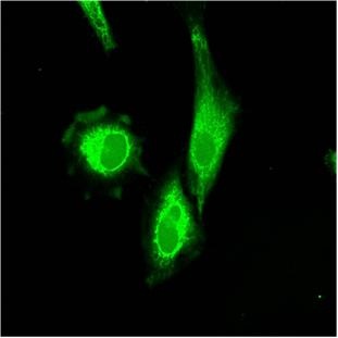
Fluorescent imaging of HeLa cell
Contact information
For more detail about research
Hirokazu Komatsu, YAMATO-MANA Program Researcher, MANA
TEL: +81-29-860-4832
E-Mail: KOMATSU.Hirokazu nims.go.jp
TEL: +81-29-860-4832
E-Mail: KOMATSU.Hirokazu
nims.go.jpFor general inquiry
Public Relations Office, NIMS
TEL:+81-29-859-2026
FAX:+81-29-859-2017
Email: prnims.go.jp
TEL:+81-29-859-2026
FAX:+81-29-859-2017
Email: pr
nims.go.jp