Success in Intracellular Imaging of Cesium Distribution in Plants Used for Cesium Absorption
Anticipated for applications in the development of "superplants" for radiocesium decontamination
2014.06.16
(2014.07.10 Update)
National Institute for Materials Science (NIMS)
RIKEN
Dr. Hirokazu Komatsu, a researcher at the NIMS International Center for Materials Nanoarchitectonics and a member of the YAMATO-MANA Program, and Dr. Katsuhiko Ariga, MANA Principal Investigator and Supermolecules Unit Director, in collaboration with postdoctoral researcher Dr. Eri Adams and unit leader Dr. Ryoung Shin of the RIKEN Center for Sustainable Resource Science, have developed for the first time a method for imaging cesium distributions in plant cells.
Abstract
- A research group led by Dr. Hirokazu Komatsu, a member of the YAMATO-MANA Program and a researcher at the International Center for Materials Nanoarchitectonics (MANA; Director General: Masakazu Aono) of the National Institute for Materials Science (NIMS; President: Sukekatsu Ushioda), and Dr. Katsuhiko Ariga, MANA Principal Investigator and Supermolecules Unit Director, in collaboration with postdoctoral researcher Dr. Eri Adams and unit leader Dr. Ryoung Shin of the RIKEN Center for Sustainable Resource Science, have developed a novel method for imaging cesium distributions in plant cells. Prior to this work, imaging of cesium distributions in plant cells had not been available.
- Since the accident at the Fukushima Daiichi Nuclear Power Plant, the discharge of radioactive cesium, especially 137Cs (half-life: 30.17 years), into the environment has become a serious environmental problem. While various decontamination methods are currently being studied, methods involving cesium absorption from soil and water by plants (phytoremediation) has drawn attention since they can be used to concentrate cesium, produce little waste, are inexpensive, and environmentally benign. Moreover, phytoremediation does not require removal of fertile surface soil, which is the current method applied for decontamination. Thus, phytoremediation has the advantage of being applicable in agricultural areas. Despite the low absorption rates of existing plants, this method promises many advantages and there are current urgent efforts being made to develop plants that efficiently absorb cesium. However, mechanisms of cesium transportation and accumulation in plant cells are largely unclear, and there is a lack of basic knowledge which is necessary for the development of appropriate plant species, including plant varietal improvement.
- The method developed in this research can be used to detect cesium carbonate particles at high resolution (micrometer-level) by using a fluorescent probe called "Cesium Green," which also enables intracellular imaging of cesium distribution. Imaging of cesium localization in cells of the general model plant, Arabidopsis, was performed. Following cultivation of Arabidopsis seedlings on a culture medium containing a high concentration of cesium carbonate, Cesium Green was applied to the seedlings, and the resulting green fluorescence observed was used to confirm the presence of cesium within the plants’ cells. Furthermore, fluorescence microscopy observations, which take advantage of the precise location-detecting properties of Cesium Green, revealed that cesium has a tendency to accumulate in the vacuoles of the cells.
- The technique developed in this research is anticipated to forward the elucidation of cesium transportation and accumulation mechanisms in plants and to enable the selection and improvement of plants suitable for application in phytoremediation. Since the methods involved here can be initially investigated using harmless non-radioactive cesium, which has the same chemical properties as radioactive cesium (in terms of detectability using Cesium Green), it is a highly versatile method that requires no special experimental facilities.
- The results of this research will be published in the U.S. chemical journal ACS Applied Materials & Interfaces.
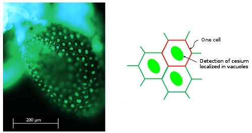
Figure 1 : Fluorescence image of Arabidopsis cotyledons (after spraying a methanol solution of Cesium Green). Bright fluorescence was observed in parts considered to be vacuoles in the cells.