Development of Coating Method that Accelerates Bonding with Bone by 3 Times
Aiming at Reducing the Burden of Orthodontic Subperiosteal Devices on Dental Patients
2013.04.08
(2013.05.22 Update)
National Institute for Materials Science
Tokyo Medical and Dental University
Dr. Masanori Kikuchi, Group Leader of the Bioceramics Group, International Center for Materials Nanoarchitectonics (MANA) and a research group at Tokyo Medical and Dental University succeeded in developing a coating method which accelerates bonding with bone by 3 times.
Abstract
- Dr. Masanori Kikuchi, Group Leader of the Bioceramics Group, International Center for Materials Nanoarchitectonics (MANA; Director-General: Masakazu Aono), National Institute for Materials Science (President: Sukekatsu Ushioda), and a research group including Masayoshi Uezona (graduate student), Prof. Kazuo Takakuda (Institute of Biomaterials and Bioengineering), Prof. Keiji Moriyama (School of Dentistry, Maxillofacial Orthognathics), and others at Tokyo Medical and Dental University (President; Takashi Ohyama) succeeded in developing a coating which accelerates bonding with bone by 3 times.
- Orthodontic subperiosteal devices are superior in terms of low invasiveness, but because bonding with bone on the surface of the bone is necessary, a minimum waiting time of approximately 3 months had been required until medical use was possible, even when coating treatment was performed with hydroxy apatite (HAp). In order to shorten this time, the device shape was optimized and a new coating method was developed in joint work by NIMS and Tokyo Medical and Dental University. As a result, a coating method which realizes in only 1 month the same bone coverage as after 3 months with the conventional device was established.
- This research result was published online on April 2, US time, in the Journal of Biomedical Materials Research Part B: Applied Biomaterials.
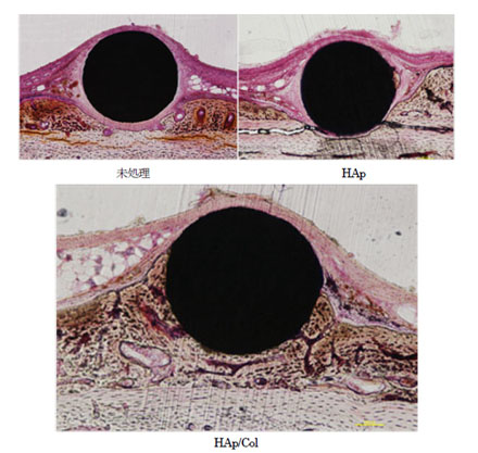
Figure : Photographs of tissue preparation during 4 weeks after surgery. In the upper 2 photos, soft tissue (dyed pink) exists between titanium material (black) and bone tissue (dyed brown); however, with the HAp/Col in the lower photos, direct bonding has occurred between the material and the bone.